The Effect of Spatial Distribution of Muscle Tissue Barrier Permeability on The Diffusion-Weighted MRI Signal
Recent simulation-based investigation of Duchenne muscular dystrophy (DMD) pathology revealed that changes in permeability have a large effect on the diffusion signal. DMD is known to cause increased permeability of muscle fibres. We use COMSOL Multiphysics® to investigate the effect of the sub-voxel patterning of permeability in muscle tissue on the diffusion signal on a model of muscle tissue. The permeability with a disordered pattern leads to statistically significant differences in diffusion signal at high sensitivity.
Simulations for diffusion MRI have historically typically employed Monte Carlo approaches; however, when we deal with exchange between compartments with different diffusivities it leads to an equilibrium condition with higher concentrations of spins in low diffusivity regions. This is a problem in (most) Monte-Carlo simulation approaches since it leads to low diffusivity regions contributing artificially highly to the overall diffusion signal, and requires an initial equilibration period which increases the computational requirements for each run. COMSOL Multiphysics® allows us to numerically solve the Bloch-Torrey equation (which is fundamental for diffusion MRI) avoiding this issue by treating the spin population as a continuum.
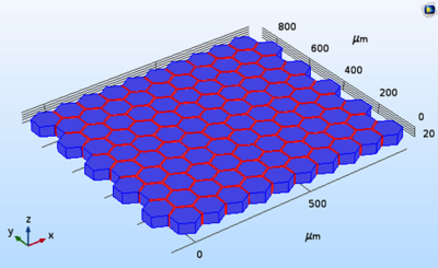
For accurate results, we require simulated diffusion to take place in an artificial environment as close as possible to muscle morphology. Muscle tissue is known from histology to be comprised of tightly-packed, non-circular fibres separated by narrow interstitial regions. As a starting point, the facilities within COMSOL Multiphysics®
allowed a hexagonal lattice with a fixed unit cell size to be set up. Cells are separated by a narrow interstitial space with a lower diffusivity. Exchange between the cells and interstitial space was via boundaries with a controlled permeability . We calculate diffusion signal as a function of scan parameters in two scenarios: one where permeability is changed uniformly across the tissue, and another in which barriers are permeable or impermeable randomly across the tissue. In both cases the total expected flux across all barriers is the same.
FEM simulations were performed using COMSOL Multiphysics® 5.4 using the Coefficient Form PDE interface from the mathematics branch. Synthetic diffusion-weighted signals were obtained by integrating the solution over the complete tissue substrate volume. Simulations of both uniform and non-uniform patterns of permeability over a range of gradient strengths and diffusion times using different intrinsic muscle diffusivities.
COMSOL® files are run on NPL’s HPC cluster to manage the 90 parameter sweep combinations. The simulations run in parallel mode with the sweep as a job array across nodes with one parameter combination per CPU.
Differences between the signal curves for the uniform and the mean non-uniform boundaries patterns became significant at long diffusion times and high gradient strengths. A two-tailed Z-test between curves showed most p-values were < 10-5 over this parameter range.
Sub-voxel distribution of permeability does lead to observable differences in the diffusion signal. The percentage differences in signals suggest that for clinically accessible scan parameter ranges, the magnitude of the effect may be too small to observe, but may become apparent on preclinical or ultra-high field human systems.