Image based, geometry prescribed CFD of intra-cardiac flows on a dynamic heart phantom
Introduction: The current gold standard for measuring intra-cardiac blood flow is cardiac magnetic resonance (CMR) and Color-Doppler ultrasound [ref1]. The presented simulation pipeline extracts heart wall motion from medical imaging and simulates the intra-cardiac blood flow as an alternative to measuring directly. The pipeline aims to compete with these direct measurement methods in two ways: 1: better spatiotemporal resolution 2: Shorter acquisition time (less breath hold, averaging over fewer heart beats)
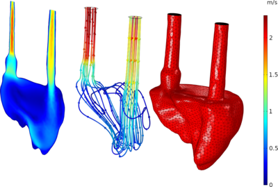
The pipeline is applied on a dynamic heart phantom for validation purposes. Materials: The dynamic heart phantom is a bi-ventricular phantom from Shelley Medical Imaging Technologies (Toronto, Canada). The phantom is mounted at the base and can be manipulated using servo motors attached to a rod at the phantom apex (compression and torsion, see figure 1). A fluid is pumped from a reservoir through an inlet of the right phantom ventricle with constant flow rate while the heart phantom is compressed 10 mm at 75 beats per minute (bpm). Methods: The dynamic heart phantom is scanned using computed tomography angiography (CTA) which provides time dependent 3D images of the anatomy. The temporal resolution is 20 volumes per heart cycle. For a heart rate of 75 bpm the resulting sample period is 0.8s/20 = 0.04s. The spatial resolution is 0.6x0.6x0.5 (mm)^3.The fluid domain is segmented from a single time instance and imported in COMSOL Multiphysics® as a surface mesh. Movement is prescribed from a displacement field obtained from volumetric image registration of all time instances i.e. all 3D frames obtained from the CTA. Therefore the displacement field has the same temporal resolution as the CTA. The segmentation and volumetric image registration is performed in MATLAB. COMSOL Multiphysics®: The heavy computational lifting is performed in COMSOL Multiphysics®. Here the surface mesh is imported and inlet, outlet and wall boundaries are defined. Moving mesh is defined for entire region and “prescribed mesh displacement” is applied to the “wall” boundary. This approach is first tested using an analytic function corresponding to the known phantom movement and later defined from the displacement field obtained from volumetric image registration. The displacement field is discrete in time and space and necessitates 4D interpolation which is implemented as an external MATLAB function. Like the phantom setup the inlet flow rate is constant 5 L/min and the outlet pressure is constant (arbitrarily set to 0 Pa). The wall has zero slip conditions where u⃗fluid=u⃗wall at the wall boundary. Results: It was possible to recreate the right phantom ventricle geometry and movement for geometry prescribed CFD in COMSOL Multiphysics®. This setup allows direct comparison with other imaging modalities such as ultrasound. The flow patterns obtained from the CFD is qualitatively similar to direct measurements using ultrasound vector flow imaging.
ref1: Intracardiac flow visualization: current status and future directions D. Muños et. al. (2013) https://doi.org/10.1093/ehjci/jet086 ref2: Validation Platform for Development of Computational Fluid Dynamics of Intra-Cardiac Blood-Flow R. Hvid et. al. (2019) https://doi.org/10.1109/ULTSYM.2019.8925893